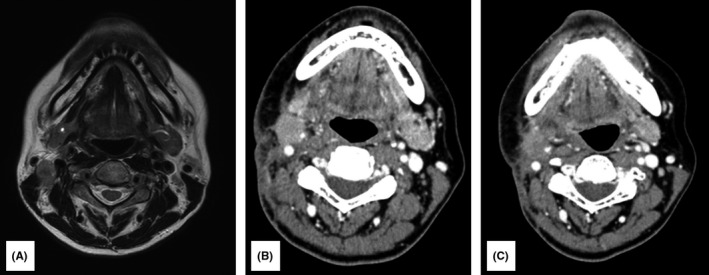
FIGURE 2.

(A) Magnetic resonance image taken 7 months after tongue cancer surgery. The right superior‐internal jugular vein lymph node had become swollen with a minor axis of approximately 12 mm, and the T2‐weighted image shows a region with moderate signal intensity that included a part of the low‐signal region. (B) Computed tomography image taken 3 months after right neck dissection. The right submandibular lymph node had become swollen with a minor axis of approximately 15 mm, and the boundary with the surrounding area was unclear. (C) Computed tomography image taken after chemoradiotherapy. No change in the size of the right submandibular lymph node was observed, and the boundary with the surrounding area was unclear
