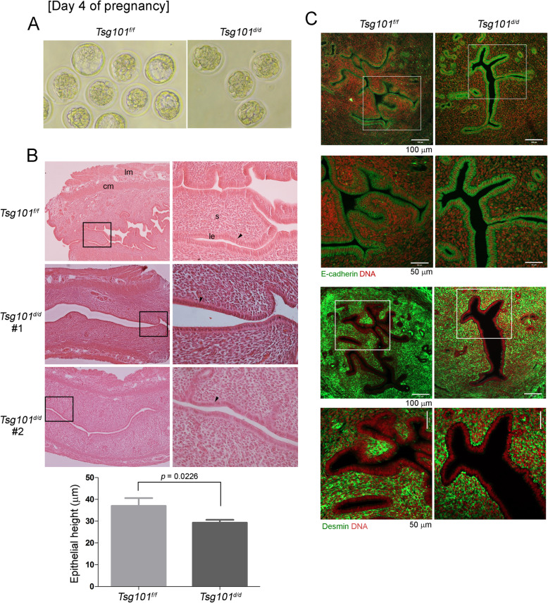
Fig. 2.
Day 4 pregnancy in Tsg101d/d mice. (A) Mice (10–11-week-old) received 2.5 IU of eCG and hCG and were bred with stud male mice. On day 4 at 11 AM, the uteri were collected and one horn was flushed with warm M16 media. A set of representative images of the retrieved embryos are shown. See Table 3. (B) Histological analysis of day 4 pregnant uterine sections. Unflushed uterine horns were used for this experiment. Paraffin-embedded sections were stained with hematoxylin and eosin. lm, longitudinal muscle; cm, circular muscle; s, stroma; le, luminal epithelium. Areas demarcated with a black rectangle are magnified in the right panels. Sections from two different Tsg101d/d uteri are shown as #1 and #2. Black arrowheads indicate the luminal epithelia. Two sections from different subjects were chosen and heights of the luminal epithelia were measured in several different areas. The measurement of epithelial heights is shown in graph. Bars represent means ± SEM. (C) Immunofluorescence staining of E-cadherin (epithelial cell marker) and desmin (stromal cell marker) was performed in one set of day 4 pregnant mice to show cell identity. Unflushed uterine horns were used for this experiment. Areas demarcated with a white square are magnified in the lower panels. DNA was counterstained with TO-PRO™-3-Iodide (1:250)

