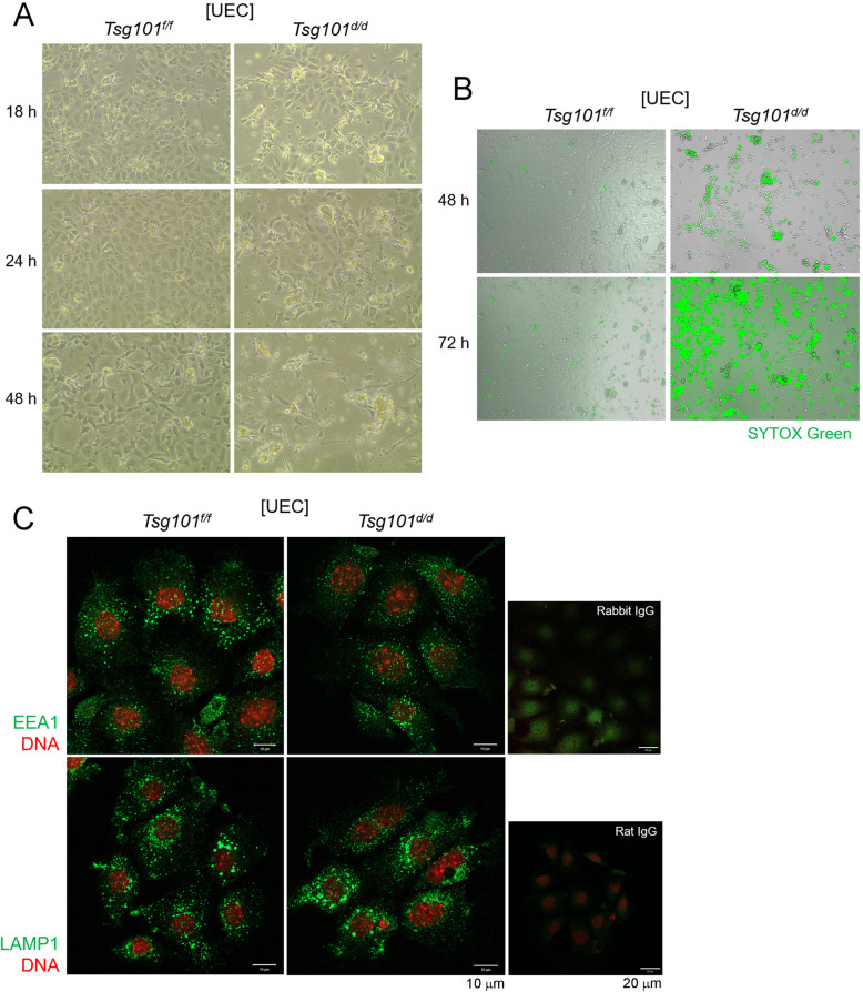
Fig. 3.
Tsg101d/d UECs gradually degenerate during in vitro culture. (A) UECs were isolated from uteri pooled from 2 to 4 Tsg101f/f and Tsg101d/d mice (11-week-old) and placed in culture at 2 × 10⁵ cells per well. An injection of E2 was administered to the mice 24 h before sacrifice to increase the cell yield. The morphology of the cultured UECs was examined at the indicated times. Experiments were repeated four times with similar results. (B) Live cell imaging of Tsg101f/f and Tsg101d/d UECs by using JuLI™ FL. 48 h in culture, cells were stained with SYTOX Green, a live dye which stains DNA of membrane-permeable cells (cells with weakened membrane or dead cells). Experiments were repeated twice with similar results. (C) Immunofluorescence staining of EEA1 and LAMP1 in Tsg101f/f and Tsg101d/d UECs. UECs isolated from Tsg101f/f and Tsg101d/d mice (9-week-old) were cultured and subjected to immunofluorescence staining 18 h later. Cells were stained with indicated primary antibodies (green). DNA was stained with TO-PRO™-3-Iodine (1:250). Experiments were repeated three times with similar results

