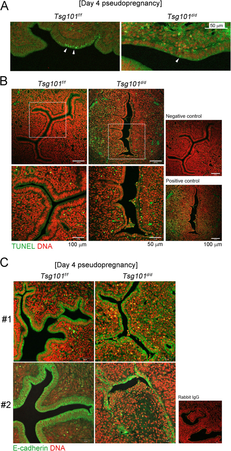
Fig. 5.

pMLKL and E-cadherin immunofluorescence staining and TUNEL assay in day 4 pseudopregnant Tsg101d/d uteri. (A) Immunofluorescence staining of pMLKL in day 4 pseudopregnant uteri (n = 2 each). One representative set is shown. (B) TUNEL staining in day 4 pseudopregnant uteri to observe apoptotic cells (n = 2 each). Green, apoptotic cell; red, nuclei. Areas demarcated with white rectangles are enlarged in the lower panel. One representative set is shown. (C) E-cadherin localization on day 4 of pseudopregnancy. Two independent samples are shown as #1 and #2 (n = 3). Uterus #2 showed the most severe phenotype of epithelial disintegration, whereas #1 showed shortened luminal epithelial height
