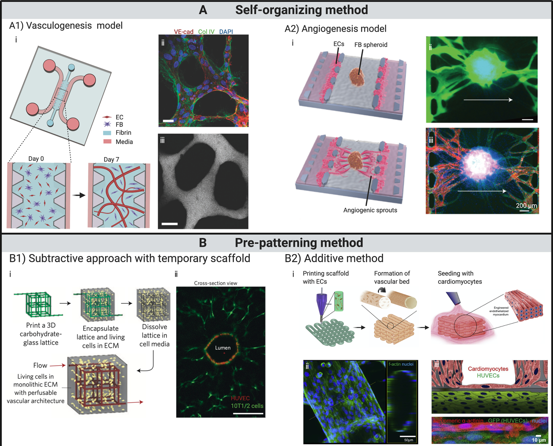
Fig 2. State-of-the-art in engineering in vitro capillary beds.

(A) Vasculature formed by self-organization. (A1) Vascular bed formed through vasculogenesis. (i) Schematics illustrate the formation of perfusable vasculature within a week by injecting mixture of ECs and fibroblasts into central gel channel of a microfluidic device. (ii) At day 7, ECs self-organized into in vivo-like capillaries with expression of continuous cell–cell junction protein VE-cadherin, and deposition of collagen IV basement membrane around the lumen. (iii) Patent lumen is confirmed by perfusion with 70 kDa dextran. Scale bars, 20 μm. (A2) Vascular bed formed through angiogenesis and subsequent integration with embedded spheroid. (i) Schematics showing fibroblast spheroid vascularized by angiogenic sprouts induced from ECs seeded on the interface of central gel channel. Perfusion within spheroid is verified with introduction of (ii) dextran dye and (iii) microbeads. (B) Vasculature fabricated through pre-patterning techniques. (B1) Functional vascular network formed around temporary scaffold. (i) Schematics showing cell-laden matrix is introduced surrounging a temporary scaffold, which is removed or dissolved afterwards. ECs are subsequently introduced into the open channels, yielding a vascular structure that matches the original scaffold. (ii) Cross-section imaging of a representative channel demonstrates endothelialized patent lumen. Scale bar, 200 μm. (B2) Vascular structures fabricated through an additive bioprinting method by directly depositing cell-laden bioinks following pre-designed patterns. (i) Schematics showing the process of fabricating endothelialized myocardium by bioprinting. (ii) Confocal fluorescent images demonstrate tubular structure formed by ECs. (iii) Schematic and confocal fluorescent image showing endothelialized myocardial tissue formed by seeding cardiomyocytes onto the bioprinted endothelialized vascular structures.
(A1) Adapted from (52), with permission of Oxford University Press.
(A2) Adapted from (23), with permission of Oxford University Press.
(B1) Adapted from (20), with permission of Springer Nature..
(B2) Adapted from (21), with permission of Elsevier.
