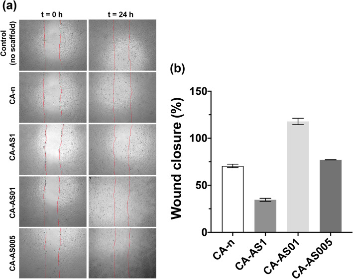
Fig. 9.
Effect of A/S mixture-containing CA scaffolds on in vitro wound healing assay using Hs27 fibroblasts. a) Microscopic images of the cell monolayer for the various samples, at 0 and 24 h after the wound was created. Red lines indicate the edges of the scratch performed in each condition. All images were recorded at 4× magnification. b) Wound closure (%) at 24 h, after treatment with the prepared scaffolds. Data are expressed as the percentage of wound closure observed in the sample where no scaffold was applied (control – 100% wound closure)

