Fig. 10.
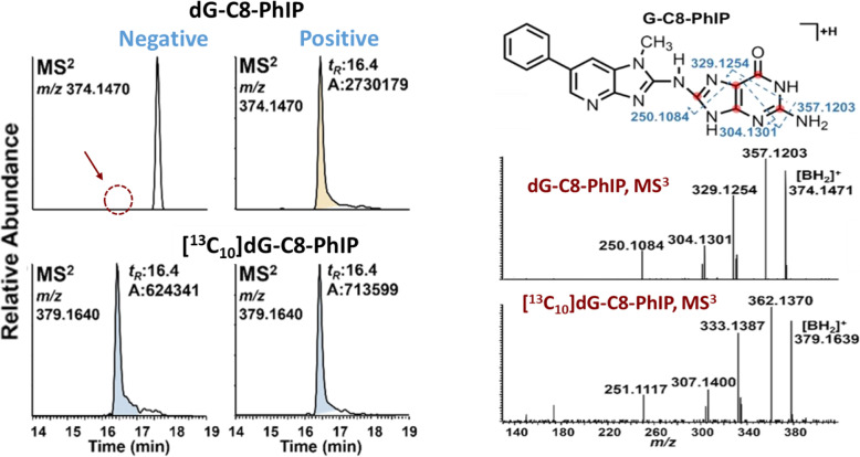
Reconstructed mass chromatograms at the MS2 scan stage of a human prostate sample targeting dG-C8-PhIP. One subject is shown with dG-C8-PhIP below the detection limit, and a second patient is positive for dG-C8-PhIP. [13C10]-dG-C8-PhIP was employed as the internal standard at a level of 3 adducts per 108 nucleotides. The MS3 scan stage product in spectra confirmed the identities of dG-C8-PhIP and its internal standard. Proposed MS3 fragmentation pathways are displayed; isotopically labeled 13C atoms of the internal standard are marked in red. Adapted with permission from [214]

