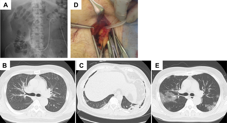
Fig. 1.
A Catheterography revealed catheter tip dislocation and omental wrapping. Outflow of contrast was limited from the side holes but not from the catheter tip. Contrast defects in the catheter were observed. B Chest CT findings on day 1 revealed bilateral pleural effusion, interlobular septal thickening, and peribronchovascular interstitial thickening. C Chest CT findings on day 4 revealed mild ground-glass opacities. D The catheter tip was wrapped by the omentum. E Chest CT findings on day 8 revealed diffuse ground-glass opacities

