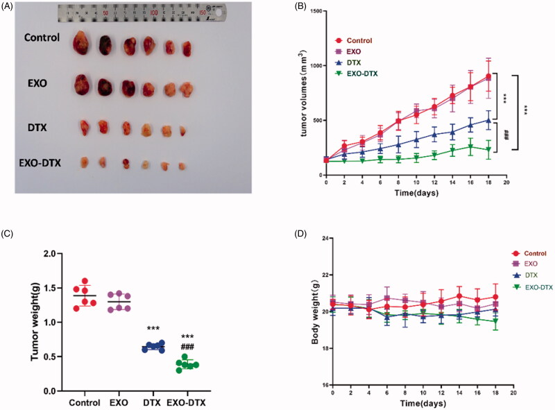
Figure 8.
Antitumor activity in vivo. Mice (n = 6) were given 19 days of oral administration of DTX, EXO, and EXO-DTX. (A) After anesthesia, tumor tissues were resected from mice for imaging purposes. (B) Tumor tissues were monitored at an interval of two days. ***p< 0.001 compared to control, ###p< 0.001compared to the DTX group. (C) Tumor weight. ***p< 0.001 compared to control, ###p< 0.001 compared to the DTX group. (D) The body weight. Values were displayed as mean ± SD. **p< 0.01, ***p< 0.001 compared to control, ##p< 0.01, ###p< 0.001 compared to the DTX group (4 mg/kg).

