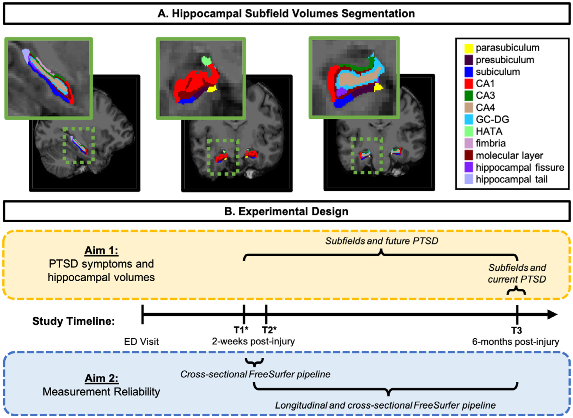
Fig. 1.

A) Hippocampal subfield segmentations from a representative participant. CA, cornu ammonis; GC-DG, granule cell layer of the dentate gyrus; HATA, hippocampal-amygdaloid transitional area. B) Schematic of experimental design including the analytic strategy for Aim 1 (yellow box) and Aim 2 (blue box) as well as the study timeline. Following the participant’s Emergency Department (ED) visit and recruitment into the study, MRI structural scans occurred at all study appointments: timepoint one (T1; two-weeks post-trauma), timepoint two (T2; two-weeks post-trauma), and timepoint three (T3; six-months post-trauma). Note: * T1 and T2 study appointments occurred on two consecutive days. (For interpretation of the references to color in this figure legend, the reader is referred to the web version of this article.)
