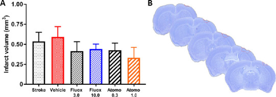
Figure 6.

Infarct location and volume.
(A) Volumetric measurements of brain infarction did not reveal statistically significant differences among experimental groups on day 42 after stroke (n = 6 per group; one-way analysis of variance followed by Dunnett’s multiple comparison, P > 0.05). Values are expressed as mean ± standard error. (B) A representative Cresyl violet-stained mouse brain on day 42 after stroke, indicating location of infarction (outlined in red dotted line) in the primary motor cortex. Atomo 0.3: 0.3 mg/kg atomoxetine; Atomo 1.0: 1.0 mg/kg atomoxetine; Fluox 3.0: 3 mg/kg fluoxetine; Fluox 10.0: 10 mg/kg fluoxetine. Similar results were observed in the second cohort of experimental animals which received high dose drug treatments but no physical rehabilitation.
