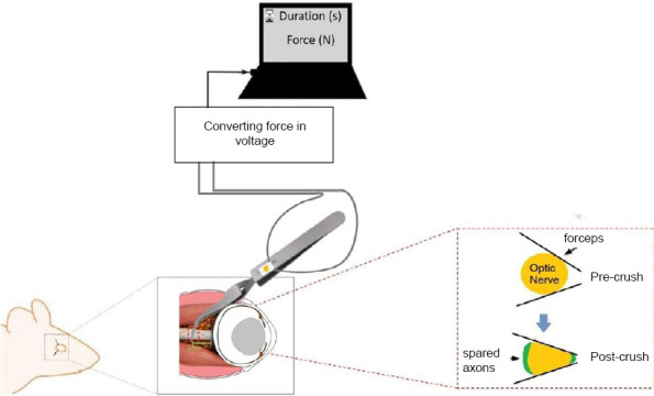
Figure 1.

Schematic illustration of the optic nerve crush (ONC) injury in mice.
The optic nerve crush is performed at about 0.5–1 mm of distance from the posterior side of an eyeball, using the instrumented self-closing #N7 tweezers. This instrument has two miniature precision foil gauges attached on each arm which could measure the force (In newton) being applied over the optic nerve. A trained experimenter can be trained with feedbacks to apply a stable and reproducible crush force. The zoom-in image of nerve crush shows the areas of spared axons over the edges of the optic nerve, due to a not perfect fit of the forceps around the nerve. See technical details in Liu et al. (2020).
