
Figure 3.
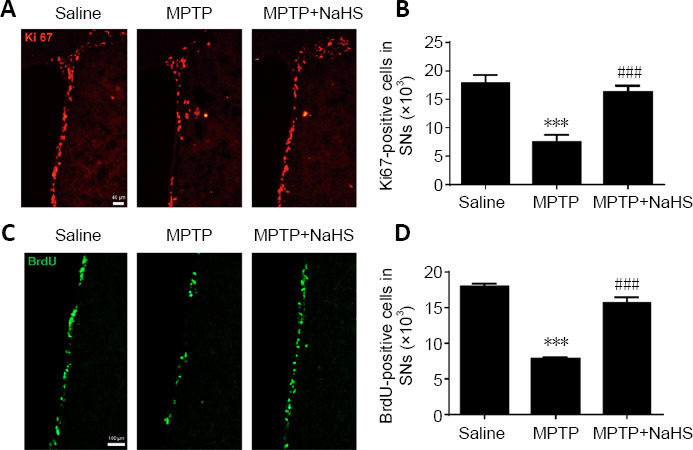
H2S promotes cell proliferation in the SVZ after MPTP-induced damage.
(A) Immunofluorescence staining of Ki67-positive (red, stained by fluorescein isothiocyanate) proliferating cells in the SVZ (original magnification 10×, scale bar: 40 μm). (B) Quantitative data for Ki67-positive cells. (C) Immunofluorescence staining of BrdU-positive (green, stained by fluorescein isothiocyanate) proliferating cells in the SVZ (original magnification 20×, scale bar: 100 μm). (D) Quantitative data for BrdU-positive cells. Data are expressed as mean ± SEM (n = 6). ***P < 0.001, vs. saline group; ###P < 0.001, vs. MPTP group (one-way analysis of variance followed by Tukey’s post hoc test). All samples were detected repeatedly in three independent experiments. BrdU: Bromodeoxyuridine; H2S: hydrogen sulfide; MPTP: 1-methyl-4-phenyl-1,2,3,6-tetrahydropyridine; NaHS: sodium hydrosulfide, a donor of H2S; SVZ: subventricular zone.