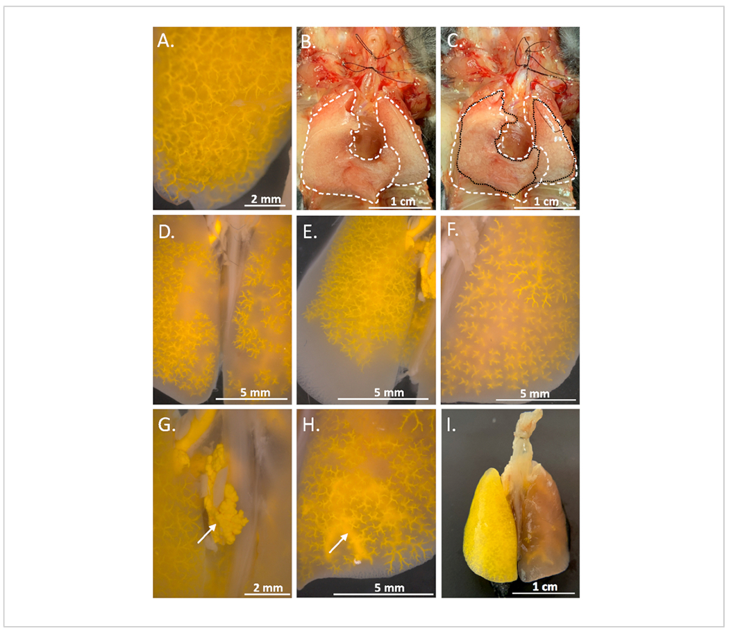
Figure 5: Examples of ideal filling and common errors during polymer compound infusion.

(A) When filling endpoint was reached, a robust and fine vascular network was observed. (B) Fully inflated formalin perfused lungs are represented by a white dashed line, (C) Underinflated/deflated lungs are shown. This was observed due to a compromised pulmonary airway. The original inflated position is represented by a white dashed line and the deflated position is represented by a black dotted line, (D) Patchy filling: the vasculature of portions of the lobe remains unfilled while other areas were entirely filled, (E) Incomplete filling: the polymer compound failed to penetrate entire sections of lung, (F) Underfilling: the polymer compound failed to fill distal vasculature, (G) Rupture: the arrow is pointing to the polymer compound extruded from vasculature, (H) Venous filling: note the arrow pointing to the arterial segments entirely filled and extending into the venous system. Veins and venules were of significantly larger caliber, (I) Catheter wedge: Here the catheter was shunted into one artery preventing the vasculature of the right lobes from filling completely while the left lobe was overfilled.
