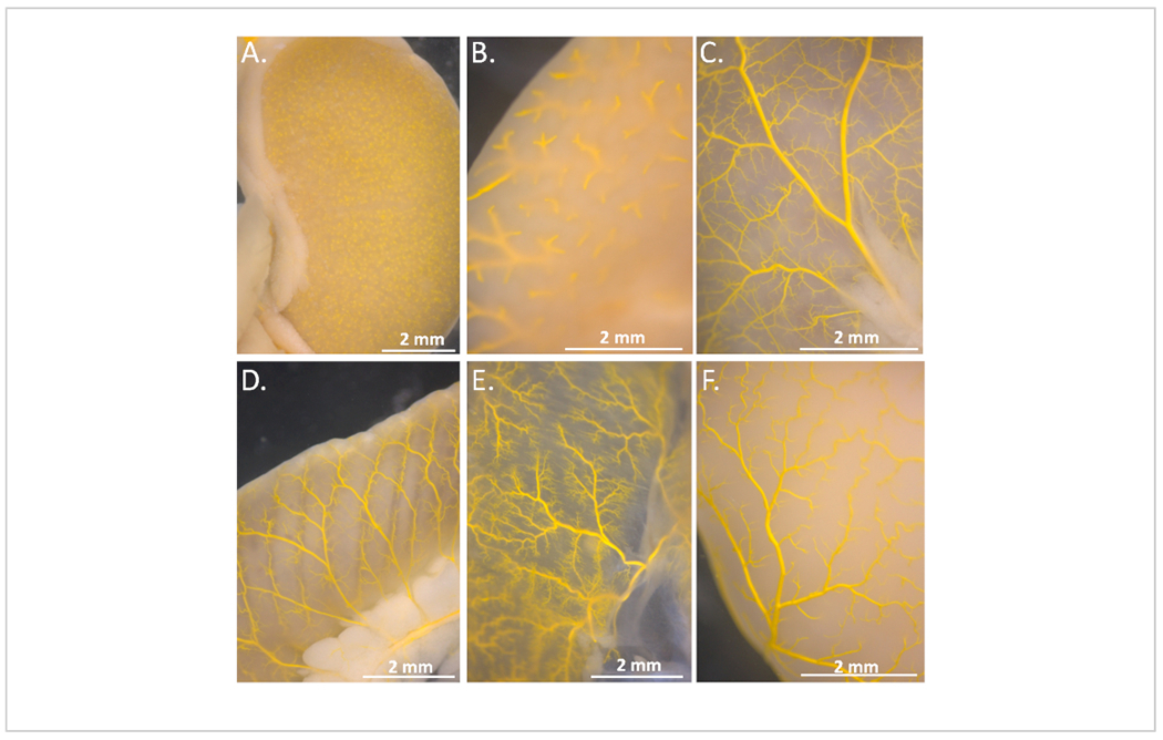
Figure 6. Vascular casting and endpoints in additional organs.

(A) Kidney: the punctate appearance of polymer compound in the glomerulus provided the endpoint. (B) Liver: note the small vessels visible at the edges of the organ. (C) Stomach: small vessels were visible and fully filled. (D) Large intestine: Small vessels are easily identifiable and filled. (E) Diaphragm: the muscle here is thin and translucent with small filled vessels apparent. (F). Brain: small vessels were visible in the cortex.
