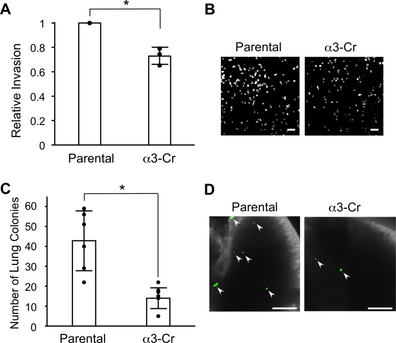
Fig 5. CRISPR-targeting of the ITGA3 gene reduces cell invasion in vitro and metastatic colonization in vivo.
Matrigel invasion assays were performed to compare invasion in parental MDA-MB-231 cells and α3-Cr cells. (A) Graph shows α3-Cr cell invasion relative to parental. Nuclei were stained with DAPI, imaged and quantified; n = 3; mean +/− SD; *p < 0.05, unpaired t-test. (B) Images show representative fields of DAPI-stained cells that invaded through transwell filters. Scale bars,100 μm. (C) Parental MDA-MB-231 cells or α3-Cr cells were labeled fluorescently by transduction with a lentivirus expressing ZsGreen, then 1 X 10⁴ cells were injected into tail-veins of 5-week old, female NSG™ mice. Graph shows number of lung colonies 21 days post-injection; n = 6 (Parental) or 7 (α3-Cr); mean +/- SD; *p < 0.05; two-tailed t-test. (D) Images show portions of lungs at time of harvest. Arrowheads point to examples of colonies detected by green fluorescence. Scale bars, 2 mm.

