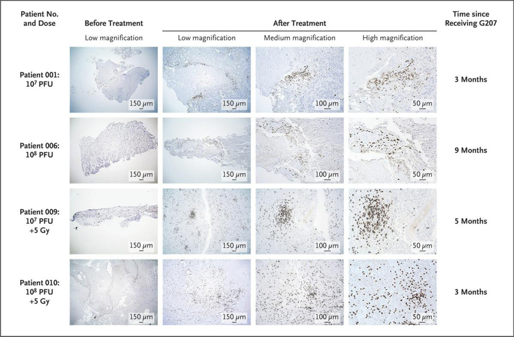
Figure 4. Immunohistologic Staining for CD8+ Cytotoxic T Lymphocytes in Matched Pre- and Post-treatment Tissue from Four Patients.
The left column shows the initial core biopsies before G207 administration; there were few immune-related cells, a finding consistent with immunologically silent or “cold” tumors. The other three columns show tumor tissues between 3 and 9 months after G207 administration from these same four patients. Post-G207 tissue revealed a brisk infiltration of CD8+ cells, which indicates an immune response to G207 and a shift to immunologically “hot” tumors. Photomicrographs were taken at low, medium, and high magnifications. Doses are in plaque-forming units (PFU) of G207 and in grays (Gy) of radiation.

