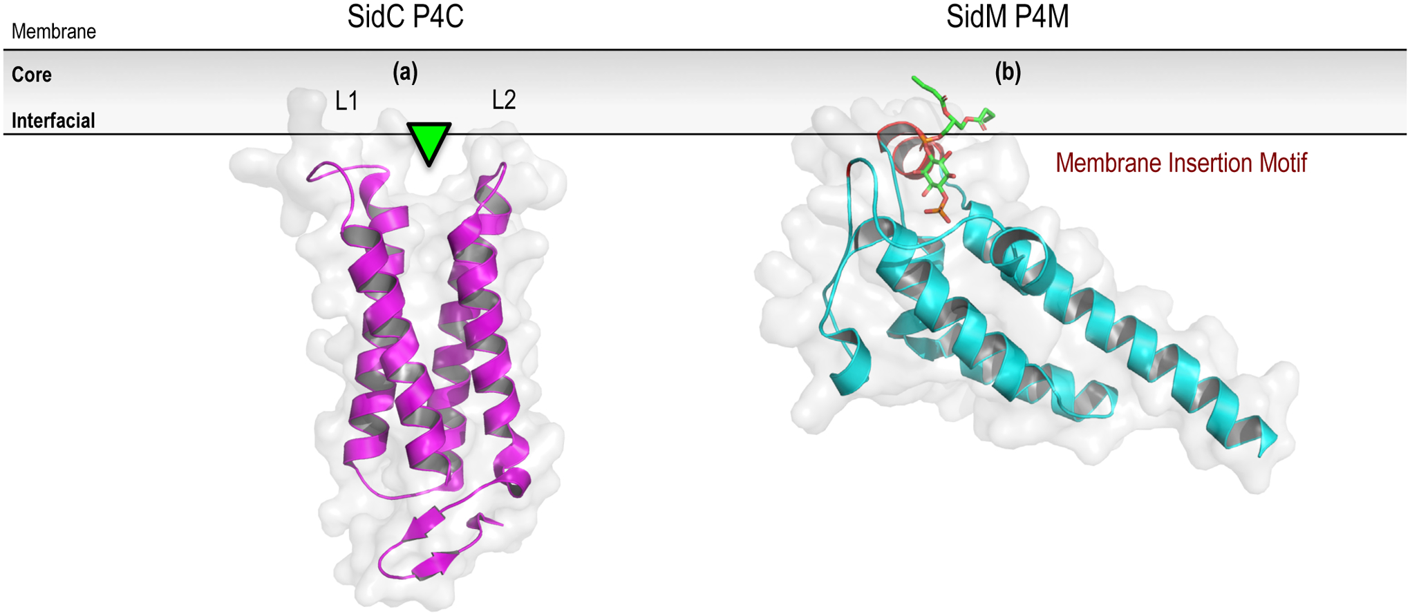
Figure 4.

Prokaryotic PPIn-binding domains. The prokaryotic PPIn-binding P4C (a; unbound structure isolated from within the full-length SidC structure; PDB entry 4ZUZ) and P4M (b; in complex with di-butyl-PtdIns4P; PDB entry 4MXP) modules are shown with their predicted membrane-bound orientations. The PPIn-binding site (green arrowhead) of the P4C has been mapped by functional and mutagenesis studies, whereas the structure of the P4M module has been solved in complex with the PtdIns4P headgroup. An elaborated membrane insertion motif that significantly penetrates the membrane, as well as contributes to the coordination of the PPIn headgroup within the binding pocket, is shown in red. For further details, please refer to Section 6 of the text. Prepared using the PyMOL Molecular Graphics System, Version 2.0 Schrödinger, LLC.
