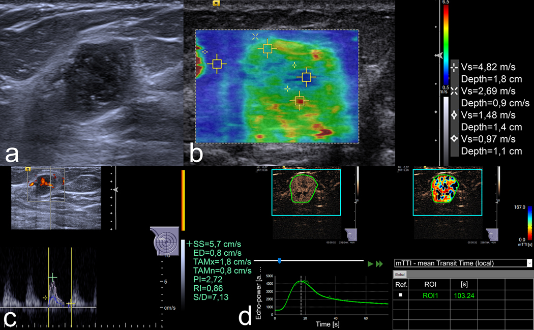
Figure 2.
US images of an 18 mm lesion in the right breast of a 54-year-old woman. (a) B-mode image demonstrates an irregular, non-circumscribed, heterogeneous lesion with posterior enhancement. Three readers classified it as BI-RADS 4b and the fourth one as 4c. (b) In VTIQ elastography, the SWVmax is 4.82 m/s. (c) Spectral Doppler shows an RI of 0.86. (d) In CEUS, the kinetic analysis shows an mTTl of 103.24 s. All quantitative parameters were above the respective cut-off values, rendering a positive mpUS. US-guided needle biopsy proved a Grade 3 invasive carcinoma of no special type.

