
Fig. 1. Foxc2 loss disrupts transport function of adult lymphatics.
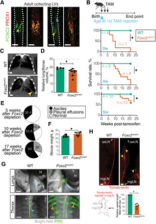
(A) FOXC2 in adult mesenteric LVs. Green, FOXC2; red, PROX1. Scale bars, 50 μm. Yellow arrowheads, FOXC2high vLECs; blue arrowhead, FOXC2+ lymphangion LECs. (B) Kaplan-Meier curve of WT or Foxc2lecKO mice survival. Tamoxifen administration at 3 weeks (*P < 0.001; n = 6 WT; n = 7 Foxc2lecKO), 4 weeks (*P < 0.01; n = 3 WT; n = 8 Foxc2lecKO), and 5 weeks of age (*P < 0.01; n = 12 WT; n = 13 Foxc2lecKO). (C) Computed tomography (CT) image of WT and Foxc2lecKO lungs. Yellow arrowhead, fluid accumulation. Scale bar, 0.25 cm. Tamoxifen administration at 5 weeks, analysis at 22 weeks. (D) Normalized WT and Foxc2lecKO lung volume. *P < 0.05; n = 4 WT; n = 10 Foxc2lecKO. (E) Ascites and pleural effusions at 5 weeks (n = 11), 10 weeks (n = 26), and 17 weeks (n = 10) after Foxc2 inactivation. (F) Increased weight of Foxc2lecKO mice. *P < 0.05; n = 10 WT; n = 9 16-week depleted Foxc2lecKO. (G) Backflow into lungs and intercostal Foxc2lecKO LVs following intraperitoneal injection of fluorescein isothiocyanate (FITC)–dextran (green). Orange arrowheads, backflow; blue arrowheads, thoracic duct (TD), lung (L), and heart (H). Scale bars, 2 mm. (H) Backflow in efferent skin Foxc2lecKO collecting vessel. Tomato-lectin injection into the inguinal LN (ingLN). Yellow arrowheads, backflow/lymph stasis. axLN, axillary LN. Scale bars, 2 mm. *P < 0.0001; n = 7 WT; n = 5 Foxc2lecKO mice. Data are means ± SD.