
Fig. 3. Foxc2 inactivation disrupts hierarchical organization of lymphatic network in an organ-specific manner.
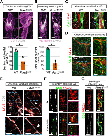
(A) Lymphatic valve leaflet degeneration in dermal vessels of 16-week depleted adult Foxc2lecKO mice ears. Pink, CD31; white, LAMA5. Yellow arrowhead, degenerated valve. Scale bars, 30 μm. Quantification of valves with mature semi-lunar shape in WT and Foxc2lecKO mice. *P < 0.001; n = 5 WT; n = 6 Foxc2lecKO. (B) Lymphatic valve leaflet degeneration in mesenteric vessels of adult Foxc2lecKO mice 16 weeks after tamoxifen administration. Staining for VE-cadherin (pink) and LAMA5 (white). Scale bars, 30 μm. Quantification of valves with mature semi-lunar shape in WT and Foxc2lecKO mice. *P < 0.001; n = 5 WT; n = 4 Foxc2lecKO. (C) Thinning of lymphatic precollecting vessels in mesentery of long-term depleted Foxc2lecKO mice. Green, LYVE1; red, αSMA; white, VE-cadherin. Blue arrow, vessel thinning. Scale bars, 50 μm. (D) Immune cell accumulation in omental lymphatic capillaries of Foxc2lecKO mice. Red, LYVE1; green, CD45. Yellow arrowhead, immune cells; blue arrowhead, LV disruption. Scale bars, 100 μm. (E) Omental lymphatic capillary disruption in Foxc2lecKO mice. Red, LYVE1; white, CD31. n = 6 WT; n = 4 Foxc2lecKO mice. White arrowheads, the point of LV disruption. Scale bars, 200 and 100 μm in magnified pictures. (F) B cells accumulate within LVs of Foxc2lecKO mice. Red, PROX1; green, B220. Blue square, accumulation of B cells within lymphatic valve sinuses. Pink square, accumulation of B cells within lymphangion. Scale bars, 50 and 20 μm for left and right images. (G) T cells around defective mesenteric lymphatic valves in Foxc2lecKO mice. Red, PROX1; green, CD3. Yellow arrowhead, defective valve. Scale bars, 50 μm.