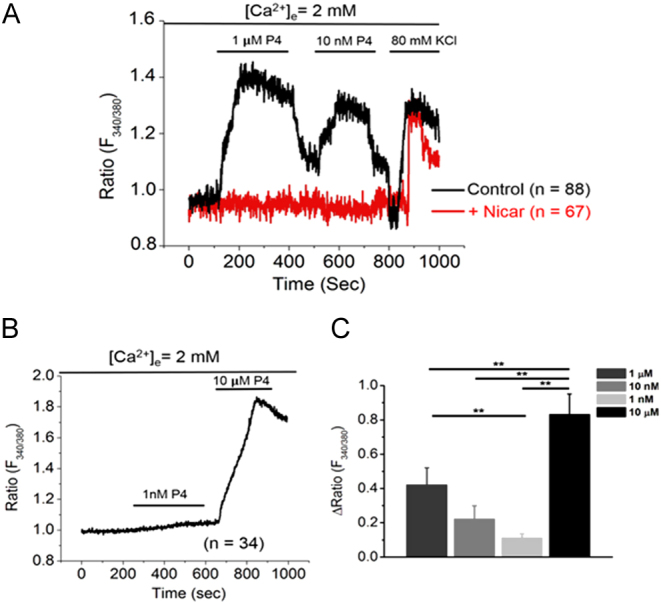
Figure 4.

Progesterone stimulates a rapid increase in intracellular calcium via L-type calcium channels in OLE cells. OLE cells were plated on 25 mm circular coverslips and incubated overnight. The following morning, cells were incubated with 2 μM Fura-2AM for 1 h at room temperature. The cells were then mounted in a flow cell with normal extracellular saline (NES) buffer. (A) Cells were treated with flow through of 1 µM P4 for 100–400 s, 10 nM P4 for 500–700 s, and with 80 mM KCl at 800 s with NES washes in between each treatment (n = 88 cells quantified). In parallel, another coverslip of cells was pretreated with 10 nM nicardipine for 30 min and then subjected to the same P4 treatment protocol (n = 67 cells quantified). (B) Cells were treated with 1 nM P4 for 100–400 s and with 10 µM P4 for 600–800 s followed by a wash at 1000 s (n = 34 cells quantified). (C) The bar graph represents the analysis of the mean change in 340/380 ratio for all P4 treatments: 1 µM (n = 88), 10 nM (n = 88), 1 nM (n = 34), 10 µM (n = 34). Statistical significance was calculated using one-way ANOVA followed by Student’s t-test. Data are expressed as mean + s.e.m. and ** represents statistical significance (P < 0.01). Analyzed data were plotted using Microcal Origin 2020 software.

 This work is licensed under a