
Fig. 4. Cytokine-producing CD4+ and CD8+ T-cells are altered by the absence of Nod2 and the cohousing conditions.
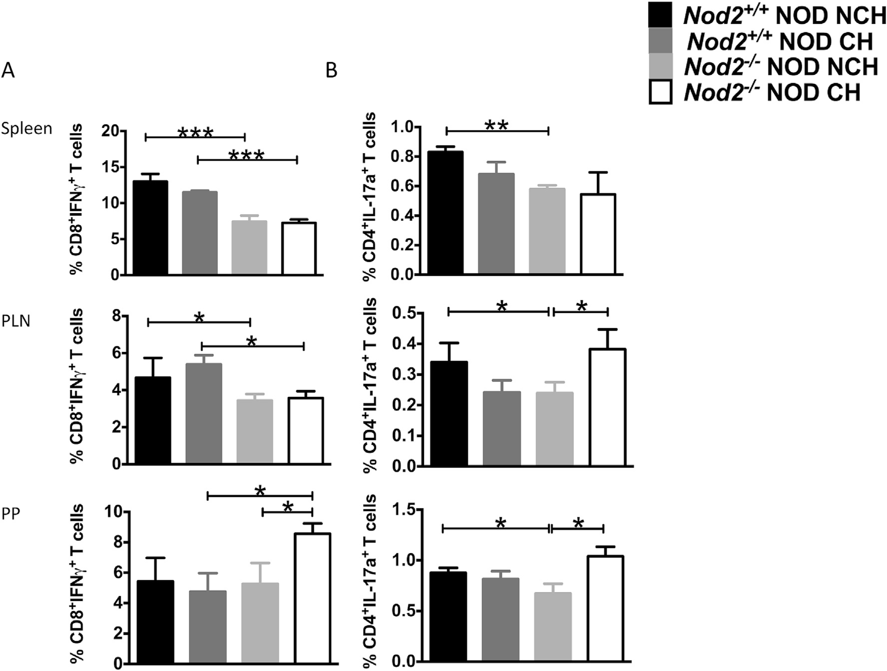
Intracellular staining was conducted on T cells from the spleen, pancreatic lymph nodes (PLN) and Peyer’s patches (PP) from 12-week old female Nod2−/−NOD (KO) and Nod2+/+NOD mice in both cohoused (CH) and non-cohoused (NCH) conditions. (A), The percentages of IFNγ-producing CD8+ T-cells and (B), IL-17a-producing CD4+ T-cells. The data represent at least 3 independent experiments (n = 3–4/group/ experiment). Student’s t-test was used for statistical analysis. *P < 0.05; **P < 0.001; ***P < 0.0001. Error bars represent SEM.