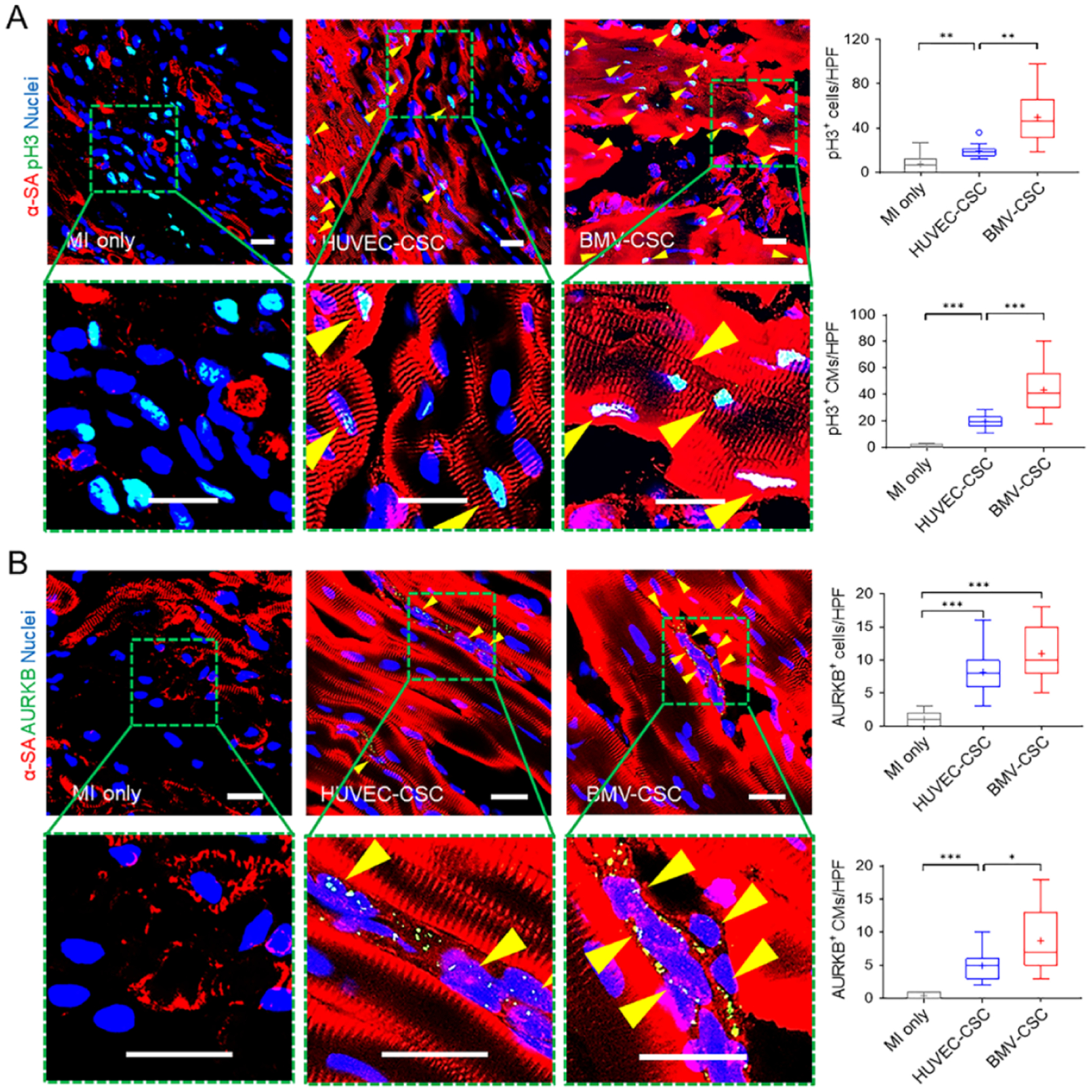
Figure 5.

BMV–CSC patch therapy promoted cardiomyocyte mitotic activity in the post-MI porcine heart. (A) Presence of pH3+ cardiomyocytes (pH3+α-SA+ double-positive cells; yellow arrowheads) in the infarcted area across different treatment groups at week 4. Quantification shows total pH3+ cells as well as pH3+ cardiomyocytes in different groups. (B) Presence of AURKB+ cardiomyocytes (yellow arrowheads) in the infarcted area across different treatment groups at week 4. Quantification shows total AURKB+ cells as well as AURKB+ cardiomyocytes in different groups (n = 3). Scale bars, 20 μm.
