
Figure 7.
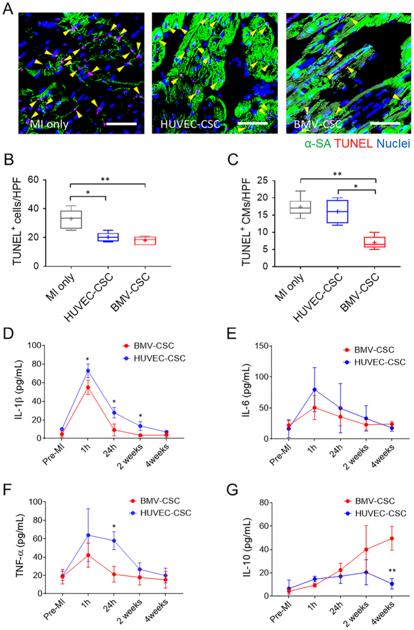
BMV–CSC Patch Therapy Suppressed Celluar Apoptosis and Inflammation in the Infarcted Porcine Hearts. (A) Presence of TUNEL+ cells in the infarcted porcine hearts across different treatment groups (n = 3) at the 4-week end point. Yellow arrowheads indicate TUNEL-positive cardiomyocytes. Quantification of TUNEL+ cells (B) and TUNEL+ cardiomyocytes (C) are performed using ImageJ software. Scale bars, 50 μm. (D–G) Porcine serum levels of IL-1β (D), IL-6 (E), TNF-α (F), and IL-10 (G) measured pre-MI, 1 h, 24 h, 2 weeks, and 4 weeks after different patch treatments, respectively. In comparison, the reference porcine serum levels of IL-1β, IL-6, and TNF-α within the first 2 weeks post-MI maintain at 100–200, 40–419, 65–80 pg mL−1, respectively.28–31