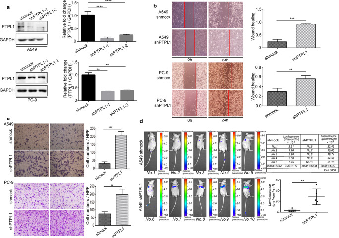
Fig. 2. PTPL1 knockdown promotes migration and invasion of NSCLC cells.
a A549 cells were infected by lentiviruses without an shRNA-expressing cassette (shmock) or those expressing shRNAs targeting PTPL1, followed by Western blot analysis of the cell lysates (each bar represents the mean ± SEM. **P < 0.01, ****P < 0.0001 vs. the shmock group). Wound healing assay (b) and Transwell assay for cell invasion (c) using A549 cells or PC-9 cells infected by lentiviruses described in (a) (each bar represents the mean ± SEM. **P < 0.01, ***P < 0.001 vs. the shmock group). d BALB-c nude mice were injected via caudal vein with luciferase-expressing A549 cells with or without PTPL1 knockdown. Bioluminescence was imaged on a Xenogen IVIS Kinetic imaging system, and the bioluminescence intensities were calculated and plotted (each bar represents the mean ± SEM. **P < 0.01 vs. the shmock group).

