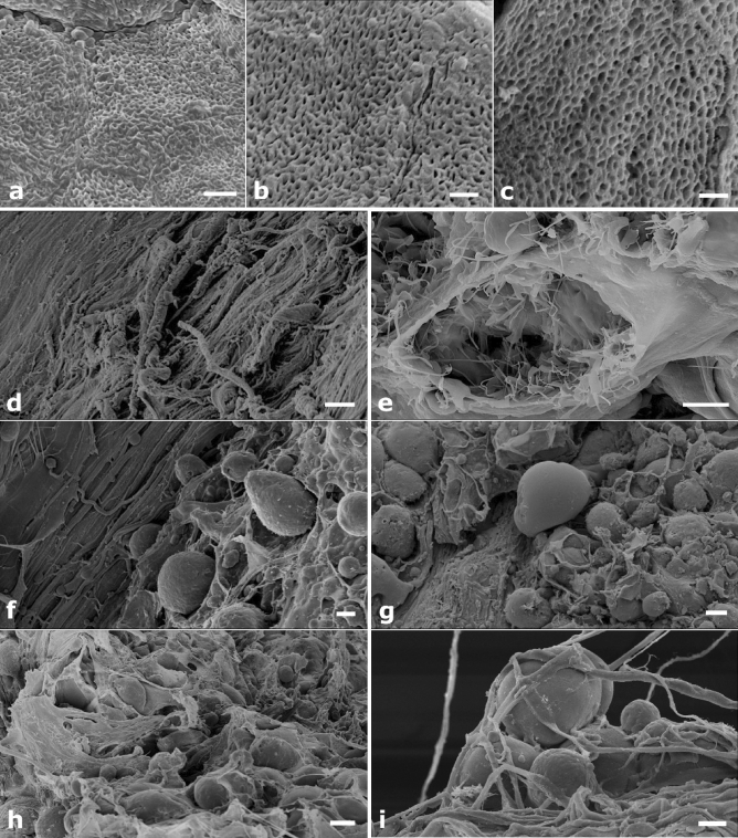
Figure 6.
(a–i) SEM images of the bone scaffold. (a–c) The tissue texture is unaltered in the natural untreated bone sample (a), it appears well preserved in the decellularized sample (b) and shows no apparent modification or damage after microporation (c). (d,e) More in detail, the decellularized and micro-pored scaffold reveals a cell-free collagen matrix (d) and well preserved cellular niches (e). (f,g) 7 days after seeding, MSCs have colonized the decellularized and pored structure, with cells randomly dispersed and well anchored to micropores (f), however they remain superficially (g). (h,i) 14 days after seeding, the scaffold is covered with cells (h) that grow inwardly in a multilayered disposition (i). Bars: (a)—2 μm; (b)—1 μm; (c)—1 μm; (d)—20 μm; (e)—5 μm; (f)—10 μm; (g)—20 μm; (h)—20 μm; (i)—10 μm.

