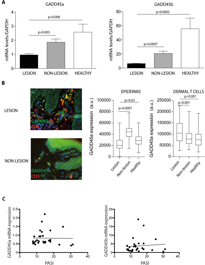
Figure 1.
Lesional skin from psoriasis patients expresses low levels of GADD45a and GADD45b. (A) mRNA levels of GADD45a (left) and GADD45b (right) were analyzed by qRT-PCR in skin samples from 30 patients with psoriasis and 15 controls. GAPDH was used to normalize gene expression. Data were analyzed by one-way ANOVA followed by Tukey’s multiple comparisons test. (B) Representative staining of GADD45a (green) and CD3 (red) in skin samples from lesional skin and non-lesional skin. Quantification of immunofluorescence staining, fluorescence intensity of GADD45 in epidermal (left) and dermal (right) infiltrating T cells was calculated using the Image J software. Differences between groups were determined by one-way ANOVA followed by Tukey’s multiple comparisons test. (C) Correlation analysis of GADD45a and GADD45b mRNA expression in lesional skin of psoriasis patients and disease severity evaluated (PASI score).

