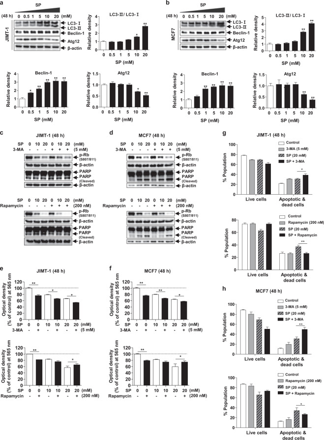
Fig. 3. Effect of SP on autophagy in breast cancer cells.
a, b Effect of SP on autophagy in JIMT-1 and MCF7 breast cancer cells. Cells were cultured with or without SP (0–20 mM) for 48 h, and the levels of light chain 3 (LC3)-II, beclin-1, and free autophagy-related protein 12 (Atg12) were assessed by Western blotting. c, d Effect of autophagic regulation on SP-induced apoptosis and proliferation in JIMT-1 and MCF7 breast cancer cells. The levels of phospho-Rb and cleaved PARP were determined by Western blotting in the cells treated with or without SP or 3-MA (autophagy inhibitor) or rapamycin (autophagy activator) for 48 h. Band densities were normalized to those of β-actin. Gel images are representative of three independent experiments. e, f The viability of the JIMT-1 and MCF7 cells treated with or without SP or autophagic regulators was measured by MTT assays. g, h Cells were treated with or without SP or autophagic regulators, and the proportions of viable and apoptotic cells were determined using a Muse® Annexin V and Dead Cell Analyzer (n = 3). Data are the mean ± SEM. Significance by Dunnett’s test: *P < 0.05, **P < 0.01, ***P < 0.001 vs. the control (no treatment group) or the indicated group.

