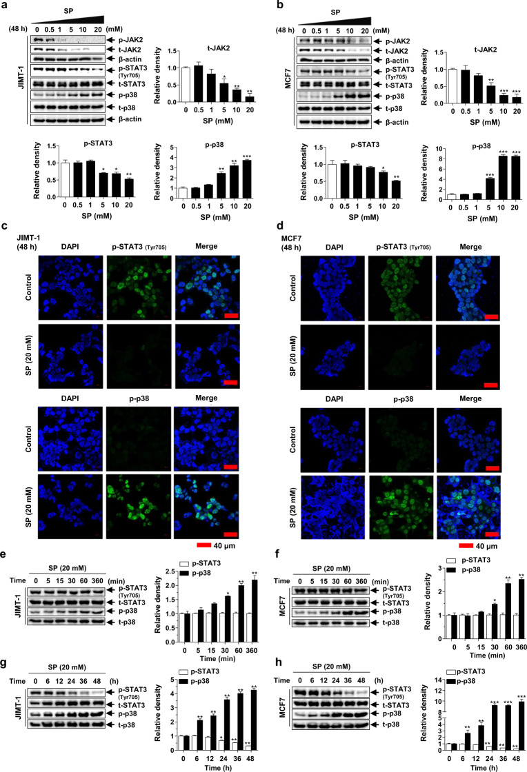
Fig. 5. Effects of SP on Janus kinase 2 (JAK2)-signal transducer and activator of transcription 3 (STAT3) and p38 mitogen-activated protein kinase (MAPK) activation in breast cancer cells.
a, b Effect of SP on the expression and phosphorylation of JAK2, STAT3 and p38 in JIMT-1 and MCF7 breast cancer cells. Cells cultured with or without SP (0–20 mM) for 48 h were lysed, and the lysates were subjected to Western blotting using the indicated antibodies. Gel images are representative of five independent experiments. c, d The cells treated with or without SP (20 mM) were incubated with primary antibodies for phospho-p38 and phospho-STAT3 (green), followed by a fluorescein isothiocyanate (FITC)-conjugated secondary antibody and DAPI (blue, nuclei). Immunofluorescence images are representative of three independent experiments (scale bar = 40 μm). The levels of phospho-p38 and phospho-STAT3 in the JIMT-1 and MCF7 cells treated with 20 mM SP for 0–360 min (e, f) or 0–48 h (g, h) were assessed by Western blotting (n = 5). The intensity of the phosphorylated protein bands was normalized to that of the total protein or β-actin. Data are the mean ± SEM. Significance by Dunnett’s test: *P < 0.05, **P < 0.01, ***P < 0.001 vs. the control (no treatment or 0 time-point group).

