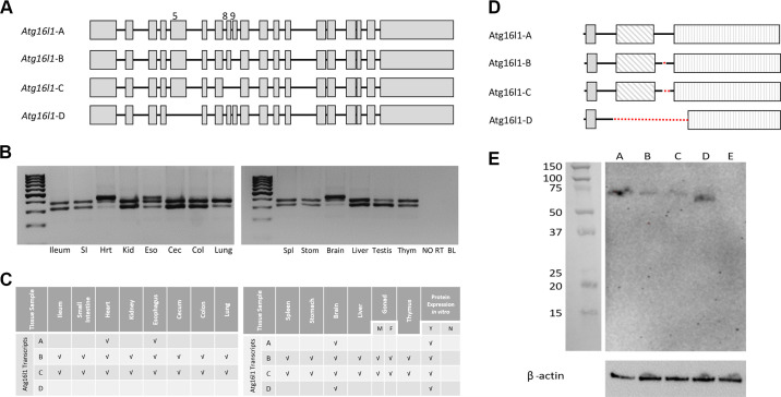
Figure 3.
Wild type (WT) splice variant DNA and protein analysis. A: representative schematic of the four WT splice variants of rat Atg16l1. Gray boxes designate exons. B: representative agarose gel images depicting splice variants detected in different tissues. C: summary of WT splice variants detected in various tissues. D: representative schematic of predicted protein isoform for each splice variant. Dotted lines denote the area of each protein isoform missing relative to the full-length protein (Atg16l1-A). shaded box, ATG5 binding motif; diagonal hashmark box, coiled coil domain; vertical hashline box, WD40 repeat domains (7 total). E: representative Western blot image showing protein expression when DNA constructs coding for each splice variant were transfected into HEK293 cells. A, Atg16l1-A; B, Atg16l1-B; C, Atg16l1-C; D, Atg16l1-D; E, nontransfected HEK293 cells (negative control). Ladder: Precision Plus Protein Kaleidoscope Prestained Protein Standards (1610375; BioRad); Loading control: β-actin.

