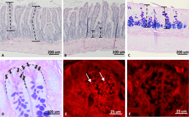
Figure 5.
Representative histologic images from intestinal characterization of the T300A rat strain. A: villus length measurement (M WT ×100; H&E). Villi were measured parallel to the center of the villus from the luminal tip to the crypt transition. B: crypt height measurement (F WT ×100; H&E). Crypts were measured from the crypt transition to the muscular layer. C: colonic mucosal thickness (M WT ×100; Alcian blue/PAS). D: Paneth cell (PC) count (M WT; ×400; Alcian blue/PAS). E and F: lysozyme IFA of ileal crypt PC. F WT, ×630 (E) and F HET, ×630 (F) HET rats exhibit inherent defects in PC granule packaging and number of granules present within the cytoplasm. Dotted lines (A–C) represent examples of how measurements were taken. Arrows (D) highlight individual PCs. Arrows (E) mark a few of many granules present. M, males; F, female; WT, wild type; HET, heterozygous.

