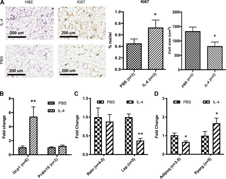
Figure 3.
IL-4 increases beige adipogenesis in neonatal iWAT. A: representative images of hematoxylin and eosin and Ki67 staining of P14 iWAT 5-µm histology sections. These images were taken at ×20 magnification and show the morphology of iWAT at P14 in PBS- and IL-4-injected cohorts and BAT at P14 in PBS-injected cohort. Ki67-stained nuclei are identified with arrows. Percentage of nuclei with Ki67 and average cell size were also quantified. qRT-PCR analysis from individual pups in 3 different litters shows the relative expression levels of beige adipocyte genes (Ucp1 and Prdm16) in P14 iWAT (n = 3–6, B), white adipocyte genes (Retn and Lep) in P14 iWAT (n = 4 or 5, C), and adipogenesis genes (Adipoq and Pparg) in P14 iWAT (n = 5 or 6, D). qRT-PCR analyses were carried out using ΔΔCt values, and data were graphed using 2−ΔΔCt (fold change of IL-4-treated samples as compared with PBS controls). iWAT, inguinal white adipose tissue. *P < 0.05; **P < 0.01.

