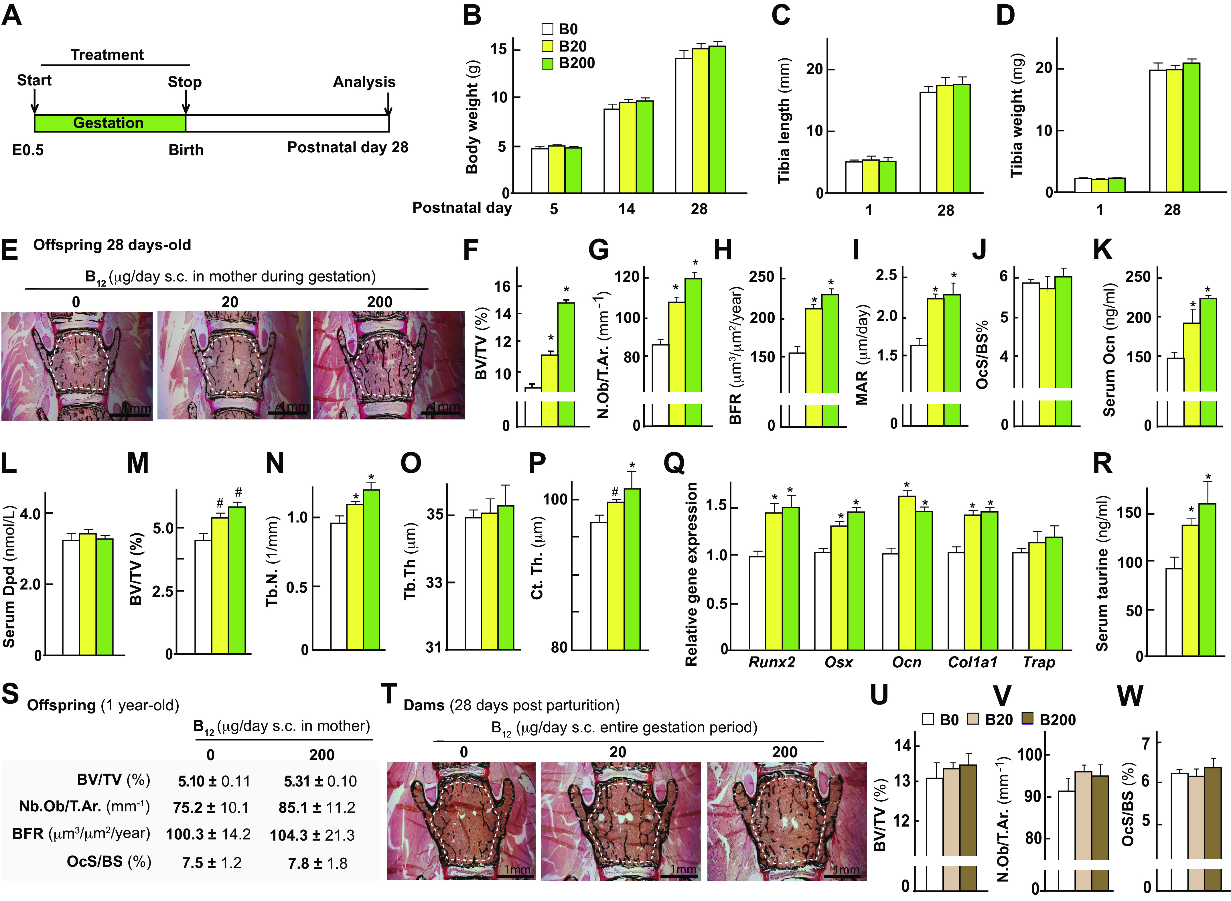
Figure 1.

Maternal B12 supplementation in the wild-type mice during gestation increases bone mass accrual of the offspring during postnatal growth. A: schematic representation of the experimental protocol used. Body weight analysis (B), tibia length (C), tibia weight (D), representative images of Von Kossa/Van Gieson-stained vertebral sections (area used to measure BV/TV% is shown) (E), bone volume over total volume % (BV/TV%) in L4 vertebral sections of the offspring (F), osteoblast number per trabecular area (G), bone formation rate (H), and mineral apposition rate (I), osteoclast surface/bone surface % (J), serum osteocalcin levels (K), serum deoxypyridinoline (dpd; L) levels in offspring from mothers that received either vehicle (0) or B12 daily through subcutaneous injections at the dose of 20 or 200 μg/day during gestation from embryonic day 0.5 to birth. Micro-CT analysis of tibia showing BV/TV% (M), trabecular numbers (N), trabecular thickness (O), and cortical thickness (P). Gene expression analysis of markers of osteoblast differentiation (Q) and serum taurine levels in offspring (R). S: histomorphometric analysis of L4 vertebra of offspring at 1 yr of age from mothers treated with either 0 or 200 μg/day dosage of B12 during gestation. Histological and histomorphometric analysis of L4 vertebra in mothers that received either vehicle (0) or B12 daily through subcutaneous injections at the dose of 20 or 200 μg/day during pregnancy: representative images of Von Kossa/Van Gieson-stained vertebral sections (area used to measure BV/TV% is shown) (T), BV/TV% (U), Osb.N./T.ar. (V), and Oc.S./BS% (W) is shown. Means ± SE is shown. n = 8–10 (A–R), n = 5 (S), n = 8 (T–W) female mice each group. *P < 0.05. #P < 0.01.
