Figure 1.
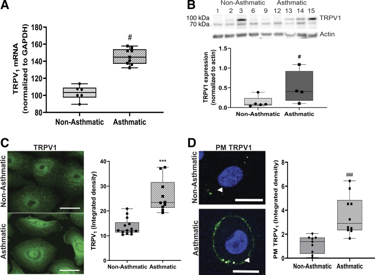
TRPV1 expression in HBE cells is greater in children with asthma versus without asthma. TRPV1 mRNA from children without asthma (1, 2, 9) and with asthma (12–15) (A) and TRPV1 protein expression in HBE cells from children without (1–3, 6, 9) and with asthma (12–15) (B) tested in two independent experiments. C: representative fluorescent micrographs of immunoreactive TRPV1 (green) localization at baseline in permeabilized HBE cells from children without (2, 6–8, 10) and with asthma (12–14) (left). Bars show the means ± SE of densitometry measurements on a minimum of 15 cells/donor (right). D: TRPV1 (green) abundance on plasma membranes (PMs) of nonpermeabilized HBE cells from children without (2–4, 8, 10) and with asthma (12, 13, 15). White arrowheads indicate the plasma membranes (left). Bars show the means ± SE of densitometry measurements on a minimum of 15 cells/donor (right). Data were analyzed using the nonparametric Mann–Whitney U test. #P < 0.05; ***P < 0.001; ###P < 0.001 compared with nonasthmatic controls. Scale bar = 20 µm. HBE, human bronchial epithelium; TRPV1, transient receptor potential vanilloid 1.

