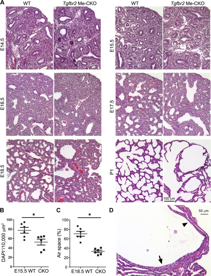
Figure 2.
Histopathology of transforming growth factor-β (TGF-β) receptor 2 gene (Tgfbr2) conditional knockout (CKO) mouse lungs. A: the hematoxylin & eosin (H&E)-stained lung tissue sections at different developmental stages are compared between Tgfbr2 CKO and wild-type mice. B: mesenchymal cell densities between embryonic day (E) 15.5 Tgfbr2 CKO and wild-type (WT) lung tissue sections are compared. Cell nuclei were detected by DAPI staining and counted in nonepithelial area. *P < 0.05 (n = 6, no. of mice). C: the peripheral (saccular) air space at E18.5, presented as the percentage of entire tissue area, was compared between Tgfbr2 CKO and WT lung tissues. *P < 0.05 (n = 6, no. of mice). D: a H&E-stained cystic structure in postnatal day (P) 1 lung of the Tgfbr2 CKO mouse, which is lined with different epithelial cells ranging from cuboidal (arrow) to flat shapes (arrowhead).

