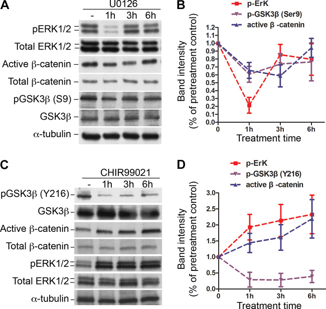
Figure 6.
ERK1/2 and GSKβ regulate Wnt canonical signaling in fetal lung mesenchymal cells. A: alterations of signal activation in primary fetal lung mesenchymal cells that were treated with ERK1/2 inhibitor U0126 (20 µM). Total cell lysates were analyzed by Western blot. α-Tubulin was used as a loading control. B: the relative intensities of the pERK1/2, pGSK3β(S9), and active β-catenin of the immunoblots in A were quantified using ImageJ and normalized with the total ERK1/2, GSK3β, and total β-catenin. C: Alterations of signal activation in primary fetal lung mesenchymal cells that were treated with GSK3β inhibitor CHIR99021 (20 μM). Total cell lysates were immunoblotted with antibodies as indicated. D: The relative intensities of the pERK1/2, pGSK3β(Y216), and active β-catenin of the immunoblots in C were quantified as described above. Compared with the untreated cells, changes of the band intensities above are all significant (P < 0.05, n = 3, no. of mice).

