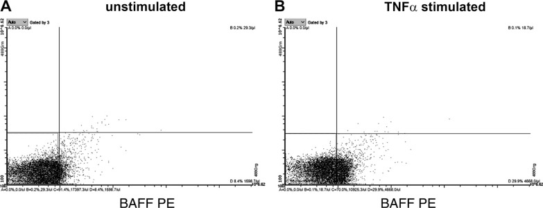
Figure 8.
Release of BAFF+ MVs from MEG01 cells is increased upon TNFα stimulation. Megakaryocyte cell line was stimulated with pro-inflammatory conditions to measure release of BAFF+ MVs. The levels of BAFF-expressing MVs increased substantially (B) after applying TNFα, compared with the unstimulated MEG01 cells (A), pointing at the role of proatherosclerotic conditions in the formation of circulating BAFF+ MVs. BAFF, B cell activating factor; MVs, microvesicles.

