
Figure 5.
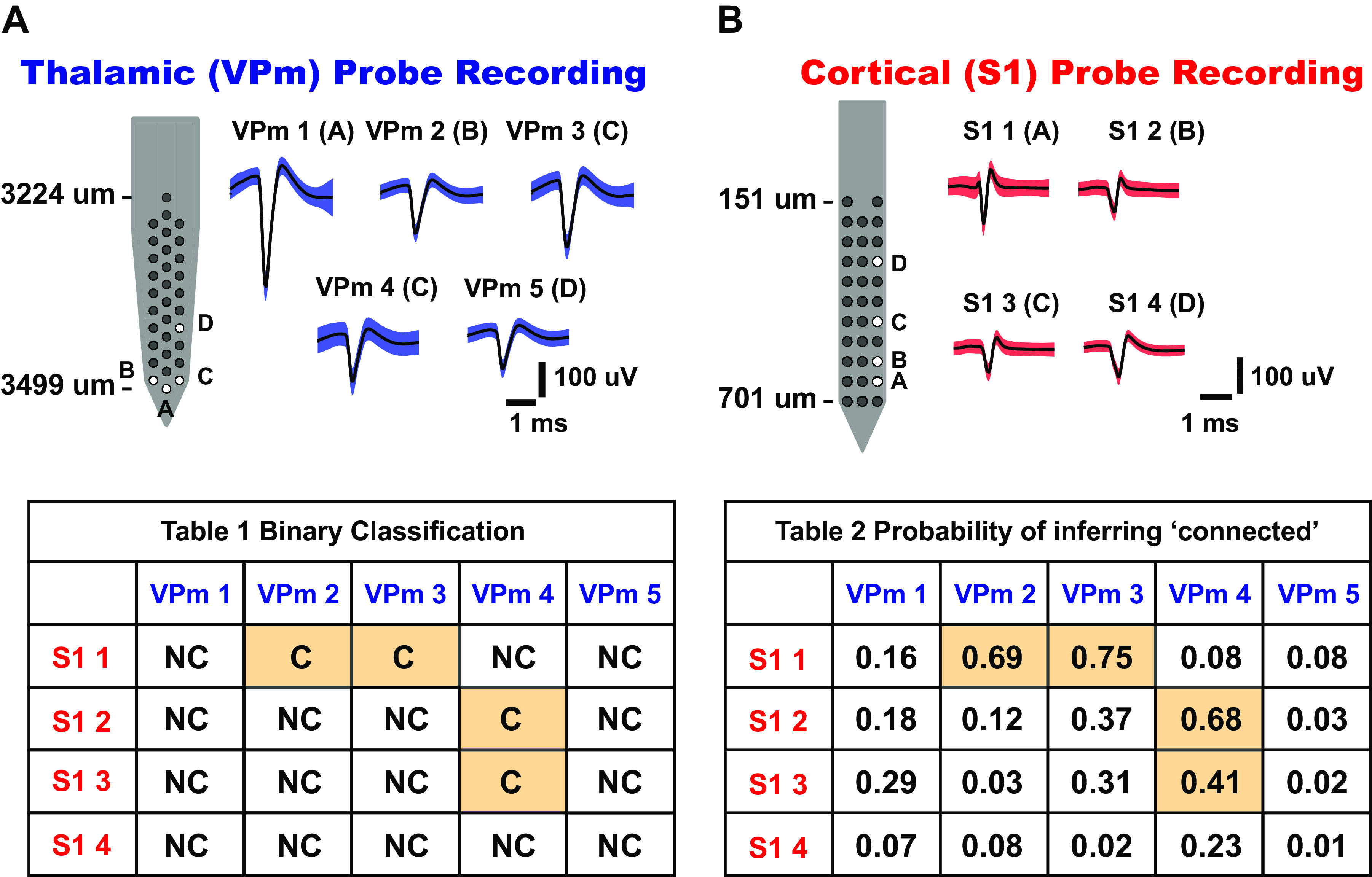
Connectivity matrix for topographically aligned, simultaneous multisite recordings. A: thalamic[ventral posteromedial nucleus (VPm)] probe recording. Five whisker-responsive thalamic units were isolated from 32-channel silicon probe sites, labeled A–D. Mean waveforms of single units are shown on right (shaded region indicates 1 standard deviation of spike amplitude) (n = 5 neurons). B: cortical [primary somatosensory cortex (S1)] probe recording. Four whisker-responsive units, putatively from layer IV barrel cortex, were isolated from 32-channel silicon probe sites, labeled A–D. Mean waveforms of units are shown on right (shaded region indicates 1 standard deviation of spike amplitude) (n = 4 neurons). Table 1 shows binary outcomes of the monosynaptic connectivity inference based on criteria 1 and 2 of cross correlation analysis. Table 2 shows the connectivity matrix tabulating the probability of inferring a putative monosynaptic connection using the bootstrapping method for each thalamocortical pair in Table 1 (bootstrap iteration = 1,000). C, connected; NC, not connected.