Fig. 1.
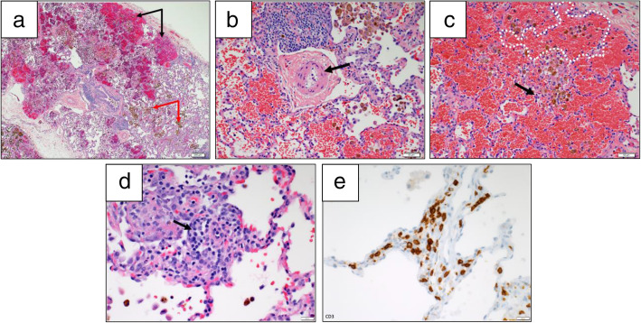
Lung biopsy findings in a 11-year-old female with trisomy 21 (Patient 3). a Low power view shows areas of alveolar hemorrhage (black arrows) and hemosiderin laden macrophages (white arrows). Hematoxylin-eosin stain, 4x. b High power view shows a moderately remodeled pulmonary artery (arrow points to pathologically muscularized arteriolar wall). Hematoxylin-eosin stain, 20x. c High power view shows hemorrhage, hemosiderin laden macrophages within simplified and distended alveoli (example is white dotted). A rare neutrophil (black arrow) is seen in the alveolar interstitium, not diagnostic of capillaritis. Hematoxylin-eosin stain, 20x. d The inflammatory infiltrate on repeat biopsy is mainly composed of lymphocytes, but rare plasma cells are noted (black arrow), 40x. e The majority of lymphocytes on repeat biopsy are marked by a T-cell immunomarker CD3, 40x

