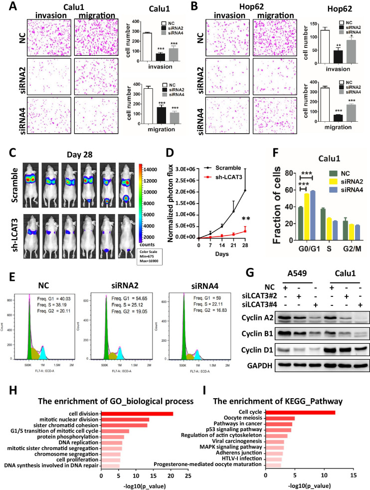
Fig. 4.
Effects of LCAT3 on migration, invasion, and cell cycle of lung cancer cells. A, B Transwell invasion and migration assays in Calu1 (A) and Hop62 (B) cells. Representative images and statistical analysis of three independent assays are shown. C LCAT3 promotes lung cancer cell metastasis in vivo. 1.2 × 10⁶ scramble or shLCAT3 cells were injected intravenously into the tail vein of nude mice (n = 6/group). The metastatic lung colonization in nude mice was measured by bioluminescence imaging. D Quantification of photon flux for metastases by tail-vein injection of shLCAT3 or scramble control Calu1 cells in nude mice. E Cell cycle analysis by flow cytometry was performed to determine the percentage of cells in different cell cycle phases. F Bar chart showing the percentage of cells in G1–G0, S, or G2–M phase. G Western blot analysis of cyclin A2, cyclin B1 and cyclin D1 in Calu1 and Hop62 cells transfected with LCAT3 siRNA or NC siRNA. H Gene ontology (GO) analysis was conducted to identify biological processes enriched for differentially expressed genes in Calu1 cells upon knockdown of LCAT3. I KEGG pathway analysis was conducted to identify pathways enriched for differentially expressed genes in Calu1 cells upon knockdown of LCAT3

