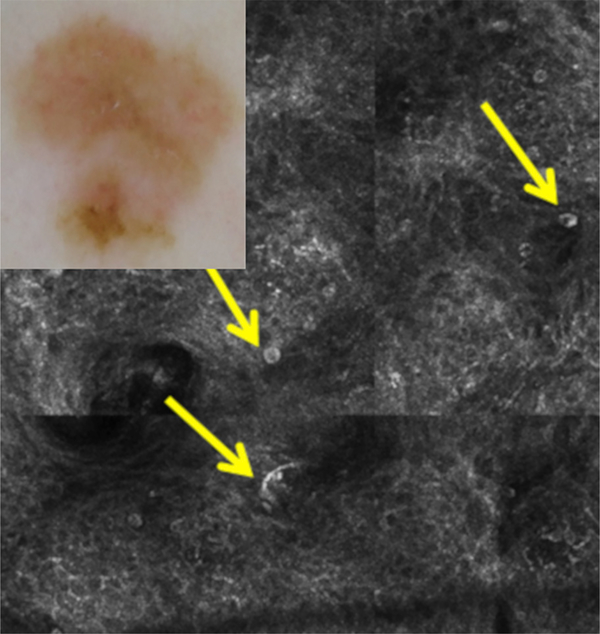
Fig 2.
A case for which RCM increased the diagnostic confidence level from 6 to 9 and changed management from observation to excision (improved diagnosis and management). Diagnosis and management before RCM: dermoscopy (inset) shows asymmetry and structureless light brown areas, nonspecific for melanoma. The clinical/dermoscopic diagnosis was atypical nevus with a confidence level of 6 (medium), and observation would have been chosen with dermoscopy. Diagnosis and management after RCM: RCM shows multiple large, round, and dendritic nucleated cells (arrows) in the superficial epidermis in a pagetoid fashion, compatible with melanoma (confidence level, 9 [high]). Excision was performed with the histopathologic diagnosis of melanoma in situ.

