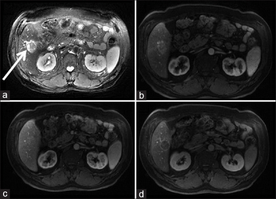
Figure 2.

Upper abdominal magnetic resonance images. T2 weighted imaging (T2WI) shows a hyperintense mass in the right lobe of the liver (a–white arrow). Early arterial enhancement has seen in contrast-enhanced dynamic T1 weighted images (T1WIC+) (b) Contrast washes out in the equilibrium phase. (c) Enhancing the capsule on the delayed images (d)
