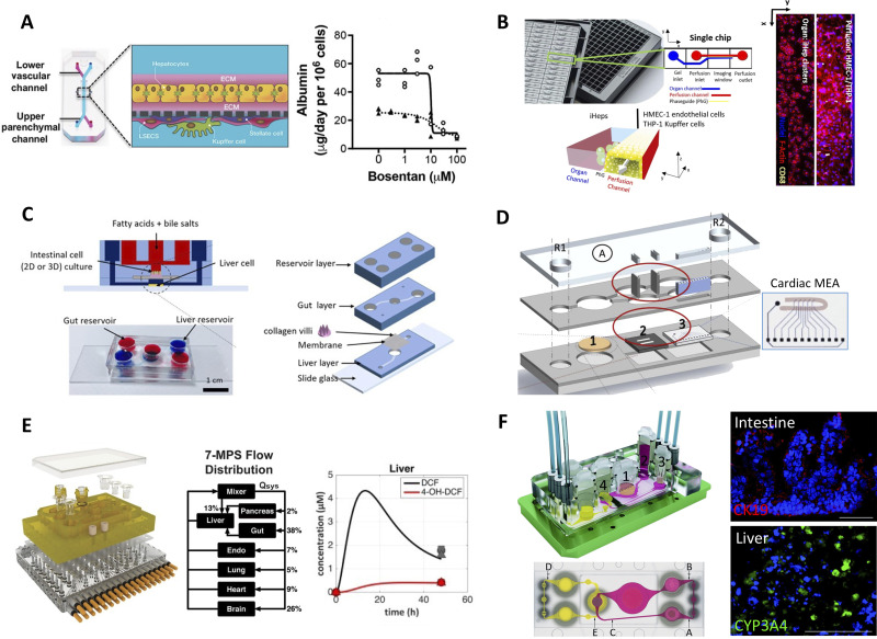
FIG. 4.
Liver-on-a-chip (microfluidic) platforms. (a) Emulate's liver-chip containing two fluidic channels separated by a porous membrane; ECM sandwiched PHHs are seeded on one side of the membrane while endothelial cells are seeded on the other side. KCs and HSCs can be added optionally to the endothelial side. Right: bosentan toxicity to PHHs in the liver-chip (solid line) and in static ECM sandwiched PHHs (dashed line).94 Reprinted with permission from Jang et al., Sci. Transl. Med. 11(517), eaax5516 (2019). Copyright 2019 AAAS. (b) Mimetas' OrganoPlate with 96 individual chips with hepatocytes seeded in the static channel and human microvascular endothelial cells (HMVEC-1) and THP-1 (monocyte line that can be differentiated into macrophages) seeded in the perfusion channel. Cell presence was verified with F-actin and THP-1 presence was verified with CD68 staining.108 Reprinted with permission from Bircsak et al., Toxicology 450, 152667 (2021). Copyright 2021 Author(s), licensed under a Creative Commons (CC-BY) license. (c) Gut–liver platform allowing for the study of fatty acid metabolism.118 Reproduced with permission from Lee et al., Biotechnol. Bioeng. 115(11), 2817–2827 (2018). Copyright 2018 Wiley. (d) Pumpless microfluidic heart-liver platform with on-chip monitoring of cardiac electrical and mechanical variations. Two laser cut acrylic (top and bottom) layers sandwich two laser cut PDMS layers with PHHs cultured on glass coverslip in chamber 1 and cardiomyocytes cultured on the cantilever array (chamber 2) as well as on the multi-electrode array (MEA, chamber 3). Medium exchange is performed through reservoirs, R1 and R2.125 Reprinted with permission from Oleaga et al., Biomaterials 182, 176–190 (2018). Copyright 2018 Elsevier. (e) Multi-MPS platform allowing for seven interconnected organ systems; each organ model on a Transwell inset can be placed into the configurable device. Right: liver compartment can metabolize diclofenac into its main metabolite, 4-OH-DCF, over time.128 Reprinted with permission from Edington et al., Sci. Rep. 8(1), 4530 (2018). Copyright 2018 Author(s), licensed under a Creative Commons Attribution (CC BY) license. (f) Multi-organ system containing small intestine (compartment 1 in schematic), liver (compartment 2), skin (compartment 3), and kidney (compartment 4) tissue models. The PDMS-glass chip in the device accommodates a surrogate blood flow circuit (pink) and excretory flow circuit (yellow) as also shown in the top view of the device (a–e indicates measurement spots in the flow circuits). Images on the right show microvilli formation (CK19 staining) in the intestine compartment and CYP3A4 in the liver compartment.131 Reprinted with permission from Maschmeyer et al., Lab Chip 15(12), 2688–2699 (2015). Copyright 2015 Author(s), licensed under a Creative Commons Attribution (CC BY 3.0) license.

