
Box 1 Figure I: 3-D structure of LC3B.
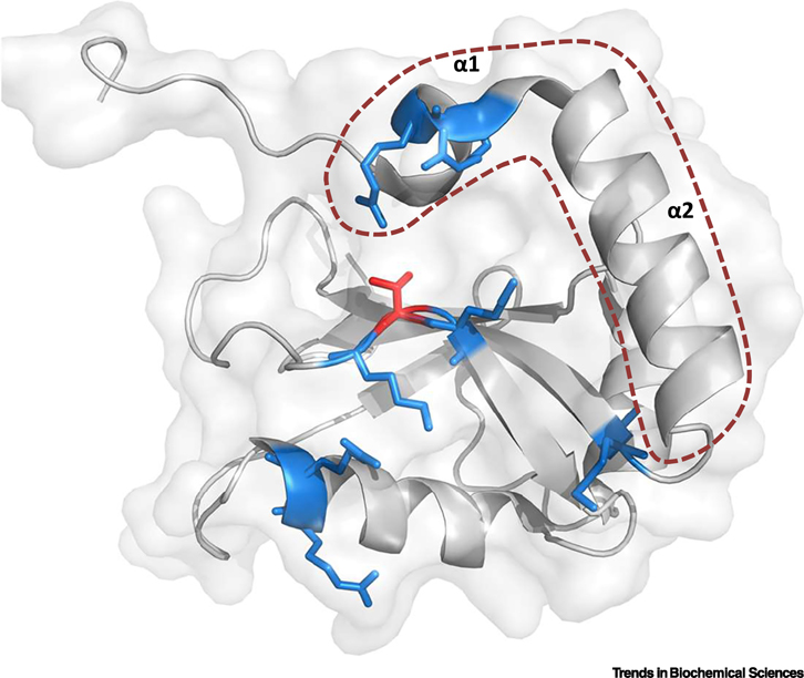
Crystal structure of LC3B showing the typical ubiquitin-like folding described for ATG8s (PDB ID 3VTU). The two consecutive N-terminal alpha helices are highlighted by an orange dotted line. Basic amino acids shown in blue form part of the LIR-docking site (LDS) and participate in the binding of LIR-containing proteins. Highlighted in red is LC3B Threonine 50, a key residue located within the LDS, whose phosphorylation regulates protein-protein interactions and is crucial for the autophagy process (see section on Regulatory mechanisms operating on ATG8 proteins that specify canonical versus non-canonical functions). Figure created with the software PyMOL.