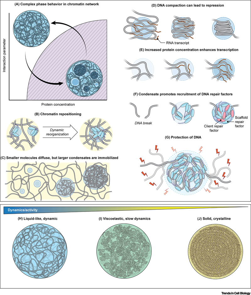
Figure 4. Regulation of phase separation in genome organization and function.
(A) Phase diagram describing phase separation of a DNA condensate within the cell nucleus. There is an interplay between organization of the chromatin and droplet coarsening. (B) Droplets nucleate around specific genomic loci, which are brought together as droplets grow and coarsen, while other genomic loci are excluded from the droplet. (C) Individual proteins and small condensates can diffuse and rearrange in the nucleus, while larger condensates are immobilized within the chromatin network. (D) Droplets concentrate DNA which may lead to transcriptional repression. (E) Droplets concentrate proteins, such as transcription factors, which can alter reaction kinetics, leading to changes in gene activity. (F) In DNA repair, droplets nucleate scaffold proteins such as 53BP1 that later recruit client proteins which partition into the droplet to augments DNA repair. (G) The viscous physicochemical environment within the droplet stabilizes stabilize the chromatin fiber from thermal fluctuations and excludes immiscible molecules from entering, such as potentially damaging reactive oxygen species (ROS). (H-J) DNA condensates exhibit a spectrum of material properties: highly dynamic, liquid-like behavior (H), intermediary, viscoelastic behavior (I), and solid-like, crystalline behavior (J). These material properties correlate with the level of biological activity, such as transcriptional bursting, controlled processing, or genome preservation, respectively.

