
Figure 1.
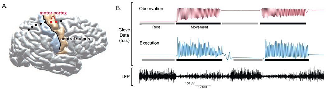
(A) Example of individual subject cortical reconstruction with electrocorticography (ECoG) electrode localization. Motor cortex recordings were obtained from bipolar electrode pair immediately anterior to the central sulcus. (B) Example glove data (top) and local field potential (LFP) recording from motor cortex during execution block. Bars below glove data indicate periods selected for rest (gray) and movement (black) periods. Movement was defined by onset and offset of clear rhythmic finger flexions. Rest periods were defined by absence of movements with brief (~1s) padding before and after rhythmic movement onset to account for transition period during which patient lifted arm from resting position. (a.u. = arbitrary units.)