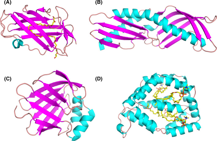
FIGURE 5.

Models of ligand‐binding proteins from less common allergen families. (A) Crystal structure of Der f 2 with polyethylene glycol (PEG; PDB code: 1XWV). While PEG originates from the solution used during crystallization, its presence reveals a large cavity that is used by Der p 2 to bind hydrophobic ligands.120 (B) Structure of Der p 7 (PDB code: 3H4Z).134 This allergen was shown to bind the bacterial lipopeptide polymyxin B. (C) Solution structure of Der f 13 (PDB code: 2A0A).168 Der f 13 is a member of the fatty acid‐binding protein family. (D) Structure of Bla g 1 in complex with lipids including phosphatidic acid28
