
Figure 5.
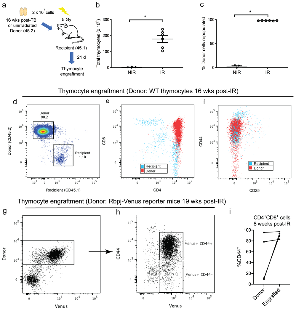
Engrafted thymocytes from irradiated donors are enriched for CD44. a, 2 x 10⁷ donor thymocytes were harvested from age matched C57BL/6J mice (CD45.2) either 16 weeks after 1.8 Gy x 4 TBI (IR) or unirradiated (NIR). Donor thymocytes were injected intravenously into B6.SJL recipients (CD45.1) 6 hours after 5 Gy TBI. Thymocytes were harvested from recipients 21 days after transplantation. b and c, The number of total thymocytes (b) and the engraftment of donor thymocytes (c) were assessed in recipients that received thymocytes from donors that were either unirradiated (NIR, n=3) or previously treated with TBI (IR, n=6). Data are presented as mean ± SE. *P<0.05 by Mann-Whitney U test. d-f, Engraftment of thymocytes harvested from wild-type mice 16 weeks post-irradiation. Representative flow cytometry plots that distinguish thymocytes derived from donor (CD45.2) vs. recipients (CD45.1). g-h, Engraftment of thymocytes harvested from Rbpj-Venus reporter mice 19 weeks post-irradiation. Donor thymocytes were injected intravenously into C57BL/6J recipients (CD45.1) Donor-derived Venus+ thymocytes were predominately CD44+. i, Transplantation of CD4+CD8+ thymocytes sorted from KrasLA1 mice 8 weeks post-irradiation. The percentage of CD44+ cells within the CD4+CD8+ population before (donor) and after transplantation (engrafted) was analyzed by flow cytometry. Each dot represents one mouse.