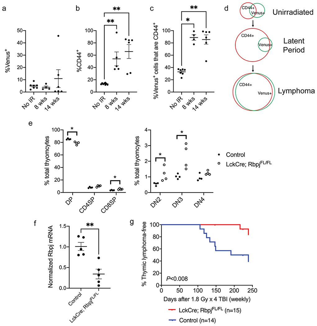
Figure 6.

Notch1 signaling is critical for the development of radiation-induced thymic lymphoma. a-b, The percentage of Venus+ cells (a) and CD44+ cells (b) in total thymocytes harvested from Rbpj-Venus reporter mice at various time points after 1.8 Gy x 4 TBI. c, The percentage of Venus+ thymocytes also positive for CD44. Data are presented as mean ± SE. Each dot represents one mouse. *P<0.05 and **P<0.01 by Mann-Whitney U test compared to No IR. d, Schematic diagram showing the changes in Rbpj-Venus+ and CD44+ cells at various stages of radiation-induced lymphomagenesis. e, The frequency of different thymocyte populations in LckCre; RbpjFL/FL mice and littermate controls without Cre. DP (CD4+ CD8+), CD8SP (CD8+), CD4SP (CD4+), DN2 (CD4− CD8− CD25+ CD44+), DN3 (CD4− CD8− CD25+ CD44−), DN4 (CD4− CD8− CD25− CD44−). n=4 mice per genotype. Each dot represents one mouse. *P<0.05 by Mann-Whitney U test. f, The expression of Rbpj mRNA in total thymocytes from LckCre; RbpjFL/FL mice and littermate controls without Cre. Data are presented as mean ± SE. Each dot represents one mouse. **P<0.01 by Mann-Whitney U test. g, Thymic lymphoma-free survival of LckCre; RbpjFL/FL mice and littermate controls that consist of RbpjFL/WT or RbpjFL/FL (No Cre) mice and LckCre; RbpjFL/WT mice that retain the expression of one Rbpj allele. All mice were exposed to 1.8 Gy x 4 TBI. P= 0.008 by log-rank test. Data were collected from at least two independent experiments.
